
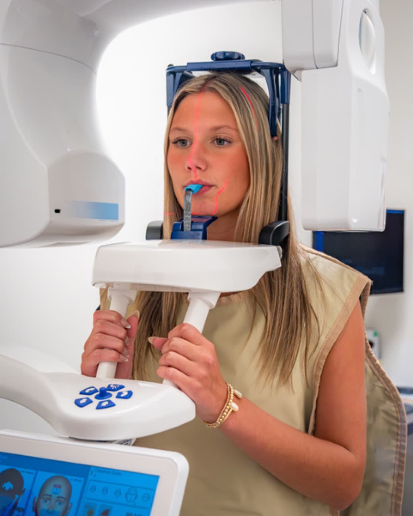
Cone Beam Imaging · Charlotte, NC
A three-dimensional view for more precise care
Some care calls for more than a flat image. Cone beam imaging captures a detailed 3D view of your teeth, jaw, and bone — the kind of precision that makes implant and complex planning safer and more predictable.
5.0 · 301 Google reviews- LVI Fellow · Neuromuscular
- Charlotte's EMFACE Provider
- 20+ Years in Dilworth
When is 3D imaging used?
Cone beam imaging is especially useful for implant planning, evaluating the jaw, and complex cases where a three-dimensional view improves accuracy and safety.
It gives us the detail to plan carefully — so your treatment is based on a complete picture.
What the scan is like
A cone beam scan is quick and completely non-invasive. You stand or sit still while the scanner rotates around your head once, capturing a three-dimensional picture in seconds — no tray, no discomfort. There’s nothing to prepare and nothing to recover from; you’re done in moments.
The result is a detailed 3D model of your teeth, jawbone, nerves, sinuses, and airway that we can rotate and measure on screen — and review with you — so complex care is planned from a complete picture rather than a flat, two-dimensional guess.
Why 3D changes planning
A traditional X-ray flattens everything into two dimensions, which can hide the exact position of a nerve, the true width of available bone, or the shape of a joint. A 3D scan removes that guesswork: for an implant, we can measure bone precisely and place with confidence; for the jaw or airway, we can see anatomy a flat image can’t show. More information up front means safer, more predictable treatment.
Who benefits most from a 3D scan
Not every visit needs a 3D scan — a routine checkup usually doesn’t. Cone beam imaging earns its place in specific situations: planning a [dental implant](/dental-implants/), where exact bone measurements matter; evaluating the jaw joint or airway; assessing wisdom teeth or an impacted tooth near a nerve; or working through a complex case where a flat image leaves questions.
When it’s recommended, it’s because the added detail genuinely improves the safety or predictability of your care — and we’re always glad to explain why it helps in your situation.
Common questions
- Its detailed 3-D view of teeth, jaw, airway, sinuses, and nerves helps evaluate TMJ disorders and joint anatomy, sleep apnea and airway narrowing, wisdom-teeth position, implant planning and bone measurement, sinus concerns, and hidden issues like root fractures or bone loss.
- Cone beam imaging uses a focused, low dose appropriate to the information needed, and we use it only when it adds real value to your care. We’re happy to explain why it’s recommended in your case.